On this page:
- Development of the Proteome Analysis Laboratory
- Proteomics, Beyond the Genome
- Proteomics Analysis Laboratory Equipment
Development of the Proteome Analysis Laboratory
The Proteome Analysis Laboratory (PAL) is a Wright State University facility for the analysis of protein/peptide expression in cells, tissues and body fluids. The goal of the PAL is to provide high-quality proteomic services to the faculty and staff of Wright State University, to researchers outside Wright State University and to provide support for intramural and extramural grants.
Mass spectrometers (MS) are at the core of the PAL. The first mass spectrometer, a Ciphergen SELDI-TOF MS, was acquired with Department of Defense grant support. It provides fast, reliable and convenient protein and proteomic profiling by using chemically treated ProteinChips® to which proteins adhere or are removed based on their specific protein chemistries. The resulting spectra can generate a proteomic profile or fingerprint of a tissue extract or body fluid (plasma or urine).
The facility was expanded in 2006 when PAL Director David R. Cool, Ph.D., received support from the Kettering Fund to purchase a High Capacity Ion Trap mass spectrometer (Bruker HCTUltra IonTrap MS). This high-end equipment allows separation of peptides and proteins on a Dionex nanoHPLC followed by nano electrospray injection into the IonTrap. Peptides are then fragmented with helium for Collision Induced Dissociation and the data processed by computer. The resulting sequences lead to specific protein identification and are matched with the proteins and peptides expected in the tissue. Post-translational modification of peptides is accomplished by Electron Transfer Dissociation.
The important next step in the development of the PAL was the acquisition of a Bruker Autoflex III MALDI-TOF/TOF mass spectrometer, through support from the Boonshoft Innovation Fund. This is a key piece of equipment for clinical and biomarker studies since it allows for high throughput screening of proteins and peptides from tissue lysates and body fluids. The increased mass resolution and ability to sequence peptides and proteins with this equipment means that it will be very useful for proteomic profiling required in clinical and animal studies.
To facilitate the separation of proteins and peptides prior to mass spectrometry, a Dionex Ultimate 3000 nanoLC system coupled to a Bruker Proteineer FC plate spotting robot has been purchased. This effectively uncouples LC separation of proteins from the mass spectrometer allowing more work to be carried out and archiving the proteins for future analysis.
In addition to the mass spectrometers, the lab has a Bectin Dickinson Free Flow Electrophoresis (FFE) system. The FFE allows for the separation of large quantities of proteins and peptides based on isoelectric point. This provides the first step in a large-scale protein separation or purification, and is similar to the first dimension of a 2D IEF gel.
Mission
The Proteome Analysis Laboratory is committed to developing cutting-edge, protein analysis techniques and protocols while expanding the foundation of scientific knowledge through developing courses and training for students, staff and faculty.
Proteomics, Beyond the Genome

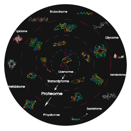
Clinical and basic research was greatly enhanced in the early 1990's by development of "gene chip" technology and subsequent elucidation of the human and other genomes. These advances expanded the ability to characterize or provide a snapshot of the genomic profile of a tissue in response to disease, pharmacological treatment or other factors. While genomic analysis provides a profile of the mRNA response, it is the proteins encoded by the mRNA that are ultimately responsible for all cellular processes and represent the endpoint response to drug treatment. Thus, the obvious next step towards understanding and monitoring the functioning of a cell or tissue in drug discovery and development is extremely complex and involves unlocking the intricate pattern of thousands of proteins expressed by the genes during the life of a cell. This approach has been given the name "proteomics" and represents tracking, sorting, characterizing and identifying the thousands of proteins in any tissue or cell to provide a fingerprint or profile of the cellular proteins under varying condition. The proteome is dynamic and can change in response to chemicals or drugs to alter the expression, function or secretion of proteins. Thus, proteomics can represent a way to initially identify diseases, potential targets for drugs and the immediate response of the cell/tissue to drug treatment. Proteomics also represents a special combination of skills and techniques that are only now becoming available and being used for comparison and characterization.
Unlike DNA and RNA, whose main purpose is to designate the function of a cell, a protein's role is to affect that function. At the basic level, protein functionality is determined by two physical parameters: 1) the protein's chemical characteristics and 2) the protein's location within the cell. The chemical characteristics of a protein are dependent on the amino acid sequence of the protein along with the secondary, tertiary and quaternary structures of the protein, as well as post-translational modifications of the protein, e.g., glycosylation, phosphorylation and enzymatic cleavages. Thus, the permutations of different proteins and peptides that can be made are far greater than the genome would predict, i.e., 20,000-25,000 genes versus 400,000 proteins.
In contrast to DNA and RNA, which are organized within specific and limited regions of a cell, proteins are ubiquitous, being found in every compartment. The function of a protein is linked to this compartmentalization. As an example, the enzyme cathepsin is localized to lysosomes where the low pH is conducive to its enzymatic activity. Likewise, receptors and transporters are found in the plasma membrane where they are responsible for transferring extracellular signals into the cell or actively chaperoning macromolecules into the cell, respectively. While this compartmentalization may complicate the process of generalizing a single protocol for purifying a protein, it is also a benefit because it can allow for the enrichment of specific compartmental fractions containing a subset of the cell's total proteome. In this way, nuclear, Golgi, ER, lysosomal, dense core granule, membrane or cytoplasmic proteins may be isolated away from the other proteins and examined more thoroughly.
Thus, the study of the cell's proteome is desirable and can be rewarding in providing intricate details on the nature of diseases and therapeutic design. To begin to develop an understanding of a cell's or tissue's proteome, specific methodologies can be established based on the chemical characteristics and subcellular localization of proteins to be studied. These methodologies can be separated into three key stages; 1) protein acquisition; 2) protein separation; and 3) protein characterization and analysis.
Proteomics Analysis Laboratory Equipment
The PAL has developed around the three stages presented above. That is, we have purchased state-of-the-art equipment necessary to conduct 'in-depth' examination of proteomes from many different sources. Most of the following equipment is available for use by booking time using the iLab web-based software system.
- BioRad BioPlex 200 Bead Based protein analysis system – This equipment allows quantitative analysis of proteins and other biomolecules based on the Luminex platform. There are more than 25 different panels including cytokines, chemokines, diabetes metabolic, cancer, apoptotic, and peptide hormone panels. Samples from humans, mice, rats, dogs, and other species are able to be analyzed with this system. Advanced analysis software is also available.
- BioRad ChemDOC MP+ with RGB LEDs Digital camera system. For western blots and fluorescence imaging.
- BioRad CX96 RealTime PCR with melt curve and other advanced software.
- BioRad NGC FPLC (Cool Lab)
- Agilent/BioTek Cytation 5 Microscope with BioSpa8 Robotic Incubator—The Cytation 5 microscope is an imager capable of automated fluorescence and brightfield analysis of live cells or fixed cells/tissue, as well as tracking of live cells over a 2 week period. It also is capable of working the same as the BioTek H1Mf plate reader described below.
- BioTek H1Mf Plate Reader—This plate reader can read absorbance or fluorescently labeled probes in a variety of plate formats. It is highly useful for enzyme assays, protein, DNA, RNA measurements, fluorescence assays and ELISAs.
- Leica DMR Fluorescence Microscope with DIC and Leica Digital Camera.
- Sorvall Discovery M120SE Ultracentrifuge
- RC5c+ High Speed Centrifuge
- Savant Automated Speed Vacuum Dryer
- FotoDyne DNA Documentation System
- BioRad Criterion 2D Dodeca Cell 2D-IEF GEL System—The BioRad 2D gel system can run up
- to 10 gels at one time in a 1D or 2D format for western blotting, fluorescent staining and scanning, and band selection for mass spectrometry.
- Fuji FLA5100—This is a high resolution (10 mm) phosphorimager/fluorimager that utilizes different laser wavelengths to analyze phosphorescent screens exposed to radiolabeled molecules, e.g., in situ hybridization of tissues, protein gels or 96 well plates. The fluorimager can scan different wavelengths of fluorescently labeled proteins or DNA/RNA in gels or on western blots, enhanced chemifluorescence.
- BioRad Experion RNA/Protein Analyzer—The Experion can analyze extremely small amounts of RNA or protein in a "chip/gel" format. The sample is compared with standards and allows both quantitative and qualitative analysis of the samples prior to running GeneChip or ProteinChip® analysis.
- Bectin Dickinson Free Flow Electrophoresis—The FFE is a "gel-less" system to separate proteins, peptides, organelles and other components of cells based on their isoelectric point (PI). This is similar to the first dimension of a 2D IEF gel, however, there is theoretically no limit to the amount of protein that can be loaded and separated. Also, the proteins are fractionated and collected in a 96 well plate from which each well containing a discrete range of proteins can be analyzed by mass spectrometery, 2D-gels, or other mechanisms.
